In-House Diagnostic
In-House Diagnostic

Biochemistry blood tests are used to evaluate the functional capacity of important organs such as the liver and kidneys. In addition to this, they are
able to detect metabolic abnormalities and measure protein, glucose, and hormone levels. On the other hand, a Complete Blood Count (CBC) is used to evaluate the three blood cell lineages : red blood cells, white blood cells, and platelets. The information derived from these tests is invaluable for the presiding veterinarian and their critical analysis, thus playing an important role in their decision making process.

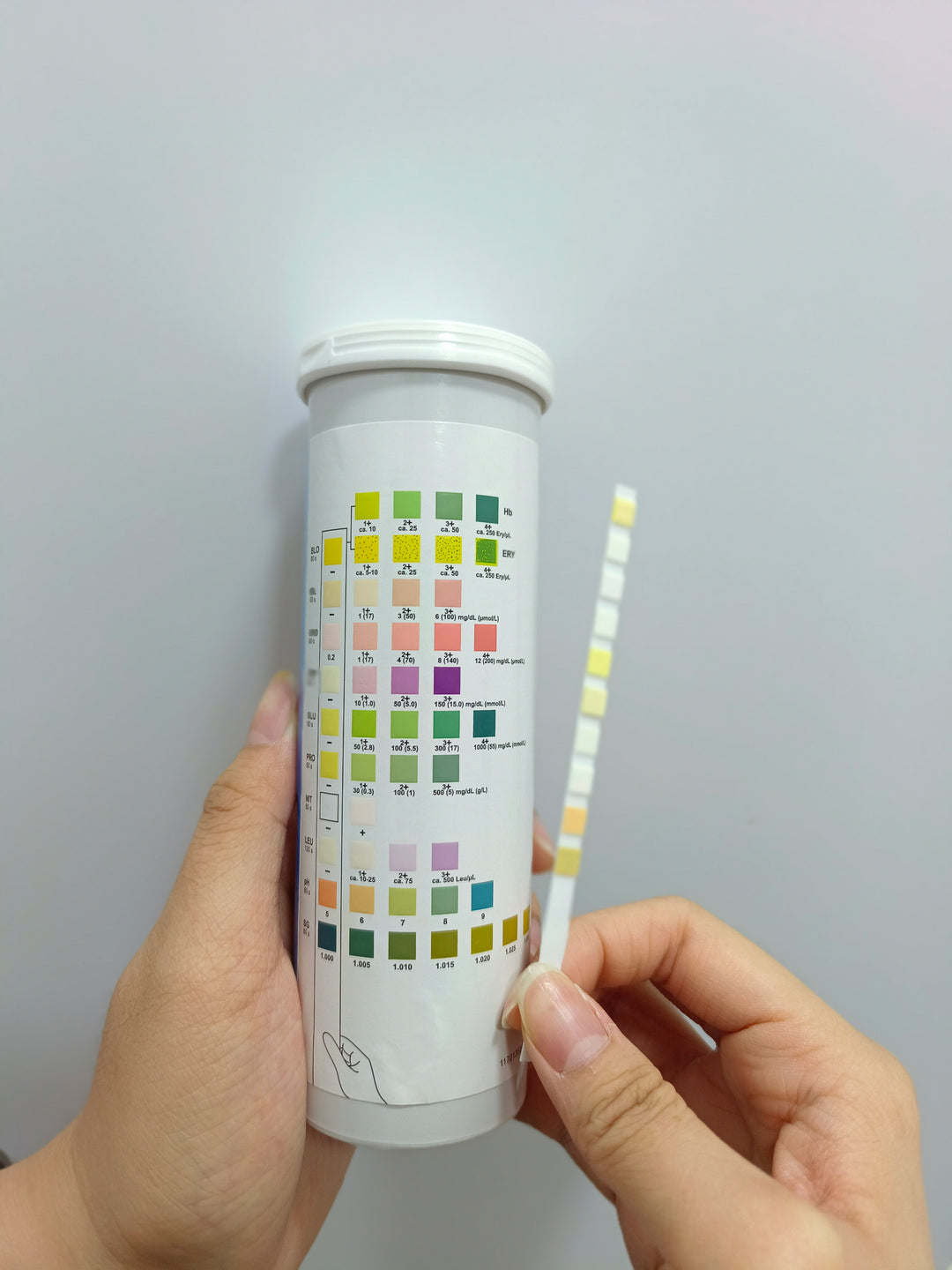
A urinalysis is a valuable indicator of hydration status and kidney function. It is also used to diagnose inflammation or infections in the
urinary tract. Microscopic examination of urine samples allows the veterinarian to observe for the presence of blood cells, bacteria, & crystals. A
dipstick analysis is likewise used to evaluate for the presence of any blood, glucose, protein, ketones, white blood cells, and urine pH.
Internal parasites can be observed and diagnosed via microscopic examination of your pet’s faeces. Often, this diagnosis is based on the presence of eggs and parasites, though their numbers and presence can be variable. As a result of this, a negative faecal analysis does not exclude the possibility of gastrointestinal parasites. At the discretion of the presiding veterinarian, further diagnostic tests may be
indicated to completely rule out the presence or absence of any parasites.

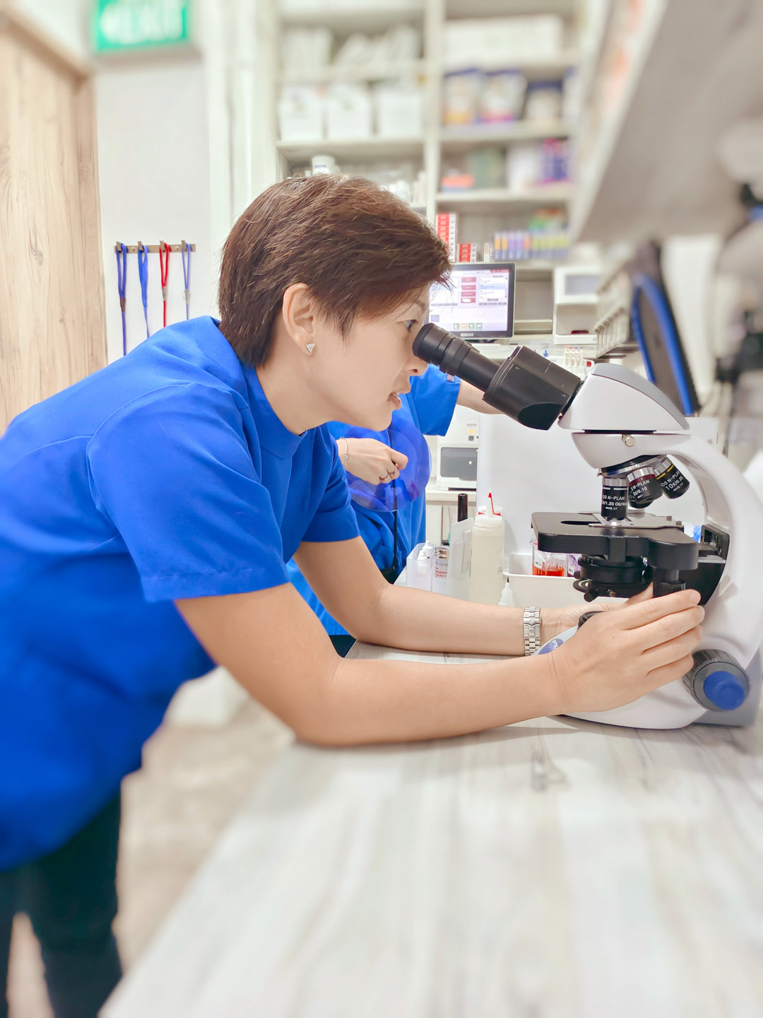
A microscope can be used to evaluate blood, urine, faeces, ear wax, other bodily fluids, parasites, cells, skin scraping and other specimens, to aid diagnosis.
CBCT (Cone Beam Computed Tomography) is a modality of advanced imaging that utilizes a cone-shaped beam of x-rays to produce detailed high resolution 3D images of structures in the skull. At Vet Central, we have successfully and reliably used CBCT imaging to diagnose and treat conditions affecting the head, neck, and oral cavity across a wide range of species. The images produced are then interpreted in conjunction with radiographic and ultrasonographic findings to reach a precise diagnosis.
While a typical scan is a rapid and simple procedure, some patients may require sedation since motion will reduce the overall quality of images produced.
Disclaimer: This video is only a demo for CBCT

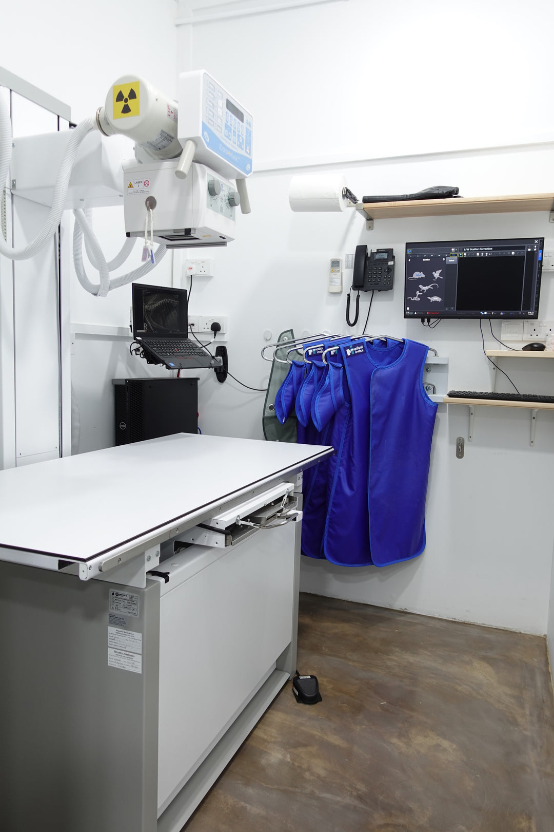
Conventional X-ray machines capture images on a film and require a higher dose of radiation to produce a clear picture. However, digital radiography uses a digital detector that is more sensitive to X-rays, allowing it to capture a clear image using a lower radiation dose. When there is injury or damage to the bone, digital radiography allows for the examination.

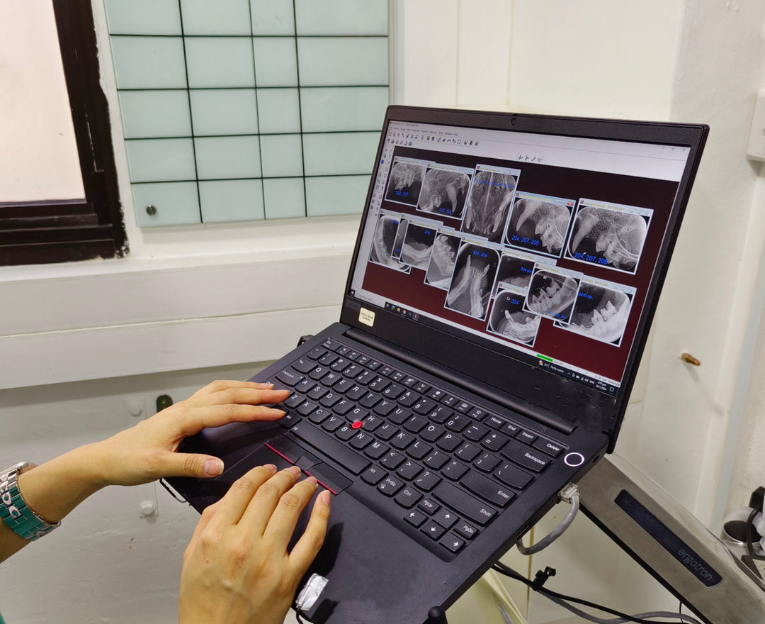
An X-ray machine uses small amounts of radiation to see the inside of your pet's teeth and areas below the gum line that are hidden from view. Unlike humans, pets must be under general anesthesia for dental X-rays.

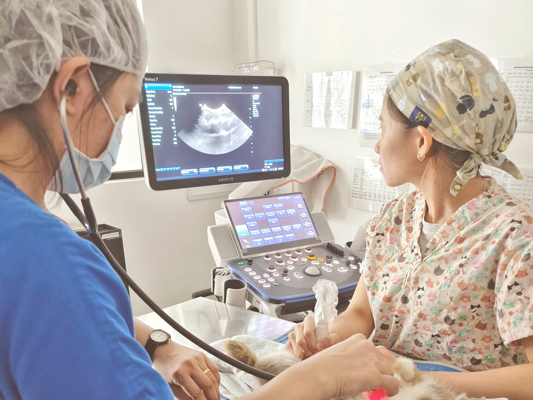
Ultrasonography is a diagnostic imaging technique that uses soundwaves to produce images of internal structures within a patient. In particular, it is used for soft tissue structures and organs such as the heart, liver, kidneys, bladder, and reproductive organs. This technique is non-invasive and generally well-tolerated by most pets. Sedation is only required in the event that a
patient is uncooperative or aggressive.
However, shaving the fur in the area of interest is necessary for clear images to be obtained. Additionally if the area of interest is in the gastrointestinal tract, fasting will be required so that a more thorough scan can be conducted.

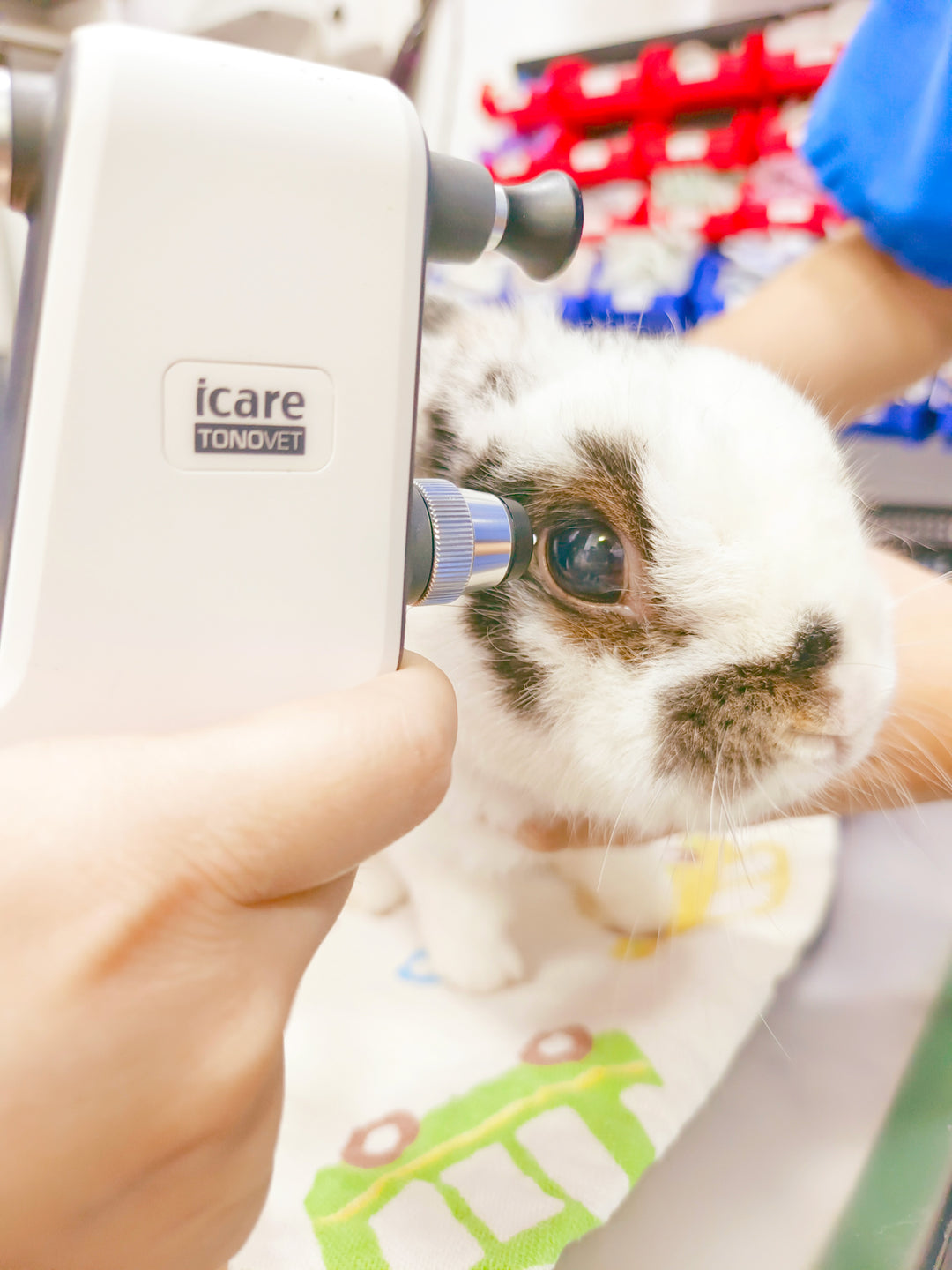
Ocular tonometry measures eye pressure and is an invaluable tool in the diagnosis of eye disorders. It is a simple procedure that makes use of a handheld device to measure eye pressure.